

Nanoparticles may be microscopic in size, but their potential in shaping imaging techniques for biomedical research is vast. Let’s explore the properties of nanodiamonds and investigate new research that is advancing their role in observing and analyzing cell processes.
A ‘Brilliant’ Solution in Biomedical Research
When scientists are interested in visualizing behavior within cells and tissues, they typically turn to organic fluorophores as a foundation for their research. Exposure to light causes these molecules to fluoresce. In other words, organic fluorophores absorb the energy from light and reemit light at a longer wavelength.
While a useful imaging technique in studying molecular processes, concerns have emerged regarding the accuracy and safety of this method. Under the illumination of light, organic fluorophores can sometimes experience degradation and bleaching with the passing of time. This not only affects their precision in measuring processes within the cells, but it can often be toxic and potentially agitate or even kill the cells.
The possibility for an alternative technique came from a rather unexpected source — nanodiamonds. Before we dive into research that has enhanced this viewing method, let’s get familiarized with nanodiamonds and learn about why they are a viable solution in biomedical research.
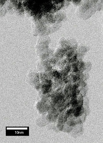
An example of a detonation nanodiamond. (“Detonationdiamond” by NIMSoffice — Own work. Licensed under Creative Commons Attribution-Share Alike 3.0 via Wikimedia Commons).
The Ins and Outs of Nanodiamonds
As their name suggests, nanodiamonds are extremely small carbon-based particles — 1,000 times smaller than a human hair, to be exact. Originally identified in meteorite impact craters, these particles share many of diamond’s valued properties, including superior hardness, chemical stability, and biocompatibility. Due to their low toxicity and compatibility with human cells, nanodiamonds have grown as a potential resource in drug delivery, serving as a carrier for transporting drugs inside cells.
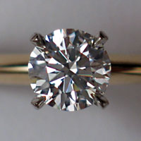
A more commercial look for diamonds. (“Diamond”. Licensed under Creative Commons Attribution 1.0 Wikimedia Commons).
Let’s pause here. Typically, when we think of diamonds, a term that comes to mind is “flawless”. We tend to place the greatest value on those diamonds that are free of defects and impurities. Ironically, in biomedical research, these defects have been a feature of value in nanodiamonds.
In previous research attempts, scientists created small defects in the crystal lattice of nanodiamonds as these flaws fluoresced when under illumination. Producing these defects, however, was a costly venture. Maintaining control throughout the process was also an issue of concern, leading to a lack of stability in the results.
This begged the question: Could there be a more effective way to view nanodiamonds in biomedical imaging? One research team sought to find out.
New Research Brings Advancements in Viewing Techniques
At Cardiff University, a team of researchers investigated a more precise and stable imaging method for nanodiamonds. Using two laser beams beating at a designated frequency, the research team created a synchronized vibration between chemical bonds in the crystal lattice structure of a nanodiamond. After producing the vibration, one beam was then used to probe the vibration and create a light known as coherent-anti Stokes Raman scattering (CARS). As the laser beams were focused onto the nanodiamond, a CARS image with high resolution was generated.
A key advantage of this technique is that it eliminates the need for adding defects to the diamonds for fluorescence purposes. Additionally, this method offers greater accuracy in measurements, as the CARS signal provided researchers with detailed information regarding the size and number of nanodiamonds delivered to the cells.
With this enhanced technique for visualizing cell processes, the potential for improved methods of drug delivery and cancer therapeutics continues to grow.


Comments (0)